
Case report | Opis przypadku
The use of split-thickness skin graft in the treatment of an atypical wound
1 Department of Wound Management, ARGO Medical Center in Lodz, Poland
2 Maria Skłodowska-Curie Provincial Specialist Hospital in Zgierz, Poland
Address for correspondence:
Gabriela Luba, Department of Wound Management, ARGO Medical Center, 27/29 Dr. Seweryna Sterlinga St., 90-212 Lodz, Poland, e-mail: lubagabriela@gmail.com
Received: 14.05.2026; Accepted: 10.08.2026
LECZENIE RAN 2026; 23 (2): 85-92
DOI: https://doi.org/10.60075/Ir.v23i2.162

Abstract
Introduction: Atypical wounds represent a heterogeneous group of ulcers characterized by unusual morphology, atypical location, lack of response to standard therapies, or pain disproportionate to wound size. Establishing an accurate diagnosis that considers the underlying etiology poses a significant clinical challenge and is essential for implementing appropriate treatment, often integrating local, systemic, and surgical approaches.
Aim of the study: To present the diagnosis and treatment process of atypical wounds using a split-thickness skin graft combined with negative-pressure wound therapy (NPWT).
Case report: A case study methodology was employed in this study. The study design based on the CARE guidelines. The present case study describes a 55-year-old female patient with chronic venous insufficiency, class III obesity, and arterial hypertension, who presented with bilateral lower extremity wounds associated with severe pain. Despite the initiation of appropriate local therapy, including hydrocolloid dressings and compression therapy, progression of the lesions and the development of satellite wounds were observed. Following the introduction of oral prednisone at a dose of 60 mg/day, inflammation subsided and wound healing improved. To accelerate healing, a split-thickness skin graft was performed. Subsequently, the wound was secured with NPWT with 80 mmHg, resulting in successful graft integration and complete epithelialization in the subsequent weeks.
Conclusions: In patients with atypical wounds, particularly those with comorbidities affecting microcirculation, consideration of an inflammatory etiology and early initiation of immunosuppressive therapy is crucial. The combination of glucocorticosteroids, split-thickness skin graft, and NPWT may constitute an effective therapeutic strategy in cases refractory to standard management.
Key words: compression therapy, negative pressure wound therapy, leg ulcer, atypical wound.
Introduction
Atypical wounds are a heterogeneous group of skin lesions that account for approximately 20% of all non-healing wounds [1]. Their etiology differs from the diabetic, venous, arterial, neuropathic and pressure-related mechanisms that underlie the classic non-healing wound. They typically show an unusual histological pattern, morphology or location, cause pain out of proportion to the size of the ulcer and fail to respond to standard treatment such as compression, optimal topical therapy and offloading of the limb [1, 2]. Recognized causes include inflammatory, neoplastic, infectious and drug-induced processes, as well as systemic disease, most often autoimmune or hematological [2, 3]. Establishing the diagnosis therefore requires a comprehensive clinical assessment, supplemented where indicated by laboratory tests, biopsy of the wound edge and imaging such as Doppler ultrasound or radiography. An atypical etiology should be suspected, and biopsy considered, whenever a wound shows no improvement after 12 weeks of appropriate standard treatment [1, 2, 4]. Because of the diversity of their etiology and presentation, atypical wounds are difficult to treat: management must rest on an accurate diagnosis and combine well-chosen topical therapy with treatment of the underlying cause. Systemic therapy is often required, including immunosuppression for inflammatory or autoimmune lesions, oncological treatment for malignant ulcers and antimicrobial therapy for wounds of infectious etiology. Optimizing nutritional status and managing comorbidities are equally essential to healing. Adjunctive therapies that promote neoepithelialization, including negative pressure wound therapy (NPWT), hyperbaric oxygen, growth factors, laser therapy and electrical stimulation, are widely used [1, 2, 5, 6], whereas bioengineering approaches based on gene therapy, 3D printing, stem cells and nanotechnology are currently the subject of ongoing research [3]. When conservative measures fail or the tissue defect is extensive, surgery may be indicated. Split-thickness skin grafting (STSG), a mainstay of reconstructive surgery, provides rapid wound coverage, stimulates healing, and is well established in the treatment of atypical wounds [1, 7]. We report the case of a 55-year-old woman with chronic venous insufficiency, class III obesity (BMI 41.0 kg/m2) and long-standing arterial hypertension. Her atypical leg wound was managed with systemic glucocorticosteroids, STSG and NPWT.
Material and methods
This single-patient case report was prepared in accordance with the CARE guidelines [8]. We describe a 55-year-old woman with progressive, painful lower-limb wounds and multiple comorbidities: chronic venous insufficiency, class III obesity and arterial hypertension. Management combined debridement of necrotic tissue with STSG and NPWT.
The patient was under the care of an outpatient wound clinic from March to May 2025. Initial diagnostic evaluation comprised history and physical examination supplemented by Doppler ultrasound of the lower limbs. Wounds were assessed with the RYB system [9], and venous disease was staged using the CEAP classification [10]. Care followed the TIMERS framework and consisted of advanced wound dressings, light compression (class 1/18-21 mmHg), debridement of necrotic tissue and pain management [11]. The wound was reassessed at every follow-up visit using the TILI score, which showed no evidence of infection [12]. When the ulcers showed no improvement, biopsy of the wound edge for histopathological examination was proposed, but the patient declined. Systemic glucocorticosteroids were therefore added, and foam dressings were used topically. After informed consent had been obtained, an autologous STSG was performed and secured with NPWT. Topical treatment, oral glucocorticosteroids and pain management were continued, and the corticosteroid dose was reduced stepwise as the wound improved, until it was withdrawn completely.
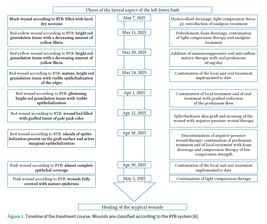
Approval was obtained from the head of the clinic, and the patient provided informed consent for publication of this case report and accompanying photographs. The study was conducted in accordance with the Declaration of Helsinki [13]. The course of treatment is summarized in Figure 1.
Case report
A 55-year-old woman presented to the wound care clinic in early March 2025 with ulcers on both lower limbs. She reported that similar lesions had first appeared in January 2025 and had initially healed spontaneously. In the weeks before presentation, however, the wounds had shown no sign of healing, the surrounding skin had deteriorated and the ulcerated area had progressively enlarged. She described severe pain in and around the wounds. Her medical history included chronic venous insufficiency, long-standing arterial hypertension and class III obesity. She had undergone surgery for varicose veins of the right lower limb several years earlier, but no operative records were available.
Arterial Doppler ultrasound of the lower limbs showed no hemodynamically significant disease, and the ankle-brachial index (ABI) was 1.05 bilaterally, confirming adequate arterail perfussion and allowing the safe aplication of compression therapy. Venous imaging demonstrated extensive neovascularization in the right groin and bilateral great saphenous vein incompetence. The left lower limb was classified as CEAP C4 Ep AS235 PR (the ulcer was not of venous origin, and the code C6 was therefore not applied).
First visit, March 7, 2025
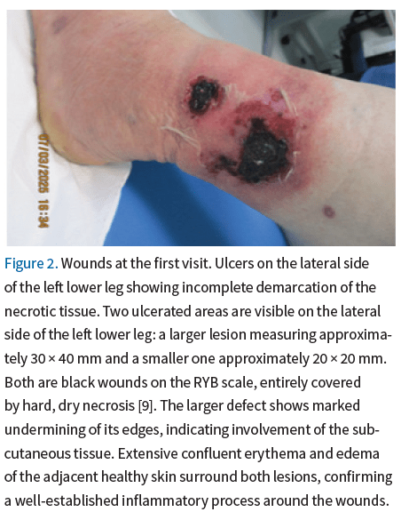
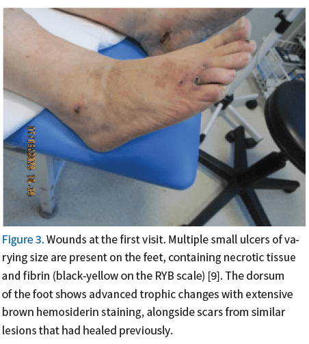
Examination showed symmetrical bilateral edema of the lower limbs, with pulses palpable bilaterally. Two wounds, measuring 30×40 mm and 20×20 mm, were present above the lateral malleolus of the left lower limb. Both were covered by hard, dry eschar, neither was exuding, and the necrosis was incompletely demarcated. The surrounding skin was erythematous, with inflammatory changes extending approximately 50 mm beyond the wound margins. Small ulcers up to 25 mm across, together with scars from similar lesions that had healed previously, were present on both lower limbs (Figs. 2 and 3).
Because demarcation was incomplete and the risk of bleeding high, sharp debridement was deferred. In line with the TIMERS framework, hydrocolloid dressings were applied to establish a moist wound environment and light compression (class 1/18-21 mmHg) was introduced. Pain management was also prescribed [5, 11].
Second visit, March 11, 2025
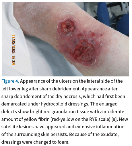
Under the hydrocolloid dressings the dry necrosis demarcated and was removed by sharp debridement. The wounds had nevertheless enlarged, new satellite lesions had appeared and the surrounding inflammation persisted (Fig. 4). Dressings were changed to foam and the remaining treatment was continued unchanged.
Third visit, March 20, 2025
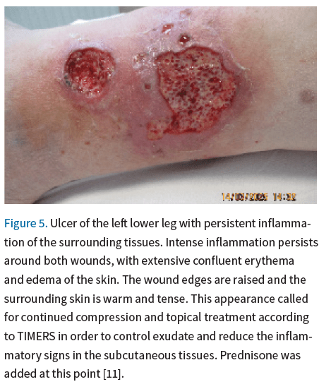
There was no response to topical treatment: periwound inflammation persisted, the ulcer beds had deepened and the satellite lesions had enlarged (Fig. 5). Pain remained severe. Given the clinical course and the atypical appearance of the wounds, vasculitis was suspected, and biopsy was planned to confirm or exclude this diagnosis. Where vasculitis is suspected the specimen must be sufficiently large, taking in the ulcer edge and, crucially, skin and subcutaneous tissue beyond the wound margin. Once the procedure had been explained, the patient declined it, fearing that the procedure would increase her pain. Immunosuppressive and anti-inflammatory treatment with oral prednisone 60 mg daily was therefore started, to be given for 10 days and then gradually reduced. Topical treatment according to TIMERS and compression were maintained [11].
Fourth visit, March 24, 2025
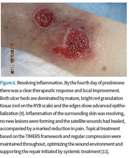
By the fourth day of prednisone the wounds had improved locally: the inflammation was settling and the satellite lesions had begun to heal (Fig. 6). No new lesions had appeared and pain had decreased markedly. Treatment was continued unchanged.
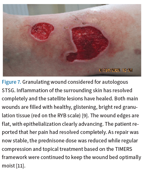
By day 11 of prednisone the inflammation had resolved and the satellite lesions were healing well. The main wounds had also improved since the previous visit, showing healthy granulation tissue and early epithelialization (Fig. 7). The patient reported that her pain had resolved completely. The prednisone dose was reduced and topical treatment was maintained. The patient was considered for STSG, which was scheduled for the following visit.
Sixth visit, April 12, 2025
On day 23 of prednisone, once the inflammation had settled, an STSG was performed in the outpatient clinic. The graft was harvested from the ipsilateral thigh with a skin graft knife; a dermatome was not used. The graft was then meshed by hand, laid over the two adjacent wounds and secured with an Avelle NPWT dressing at a continuous negative pressure of 80 mmHg (Fig. 8). The donor site was covered with a hydrogel dressing.
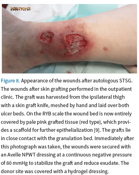
Seventh visit, April 18, 2025
On day 29 of prednisone and day 6 after STSG, the graft showed satisfactory integration, with no signs of infection (Fig. 9). The NPWT dressing was removed. Prednisone was continued at a reduced dose, together with foam dressings and low-compression bandaging.
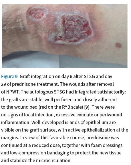
Eighth visit, April 30, 2025
On day 41 of prednisone and day 18 after STSG, the graft had integrated completely and the wounds were almost fully epithelialized, with no inflammation in the wounds or the surrounding skin (Fig. 10). Advanced dressings and compression were continued, and prednisone was reduced further as planned until complete withdrawal.
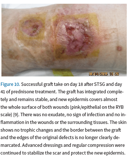
Ninth visit, May 5, 2025
Two months after presentation and 23 days after STSG the wounds were almost completely healed (Fig. 11). Light compression was maintained.
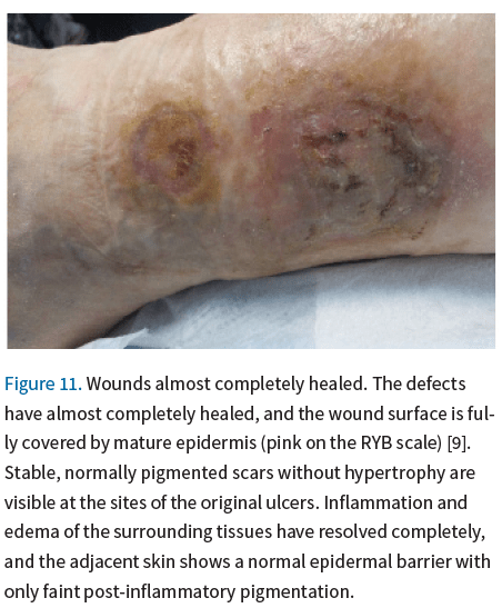
At the 3-month follow-up after the completion of treatment anf full prednisone withdrawal, the scars remained stable with no signs of wound recurrence or local inflammation. The patient continued regular prophylactic compression therapy with using class 1 (28-21 mmHg) compression garments with good tolerance.
Discussion
In this patient, chronic venous insufficiency due to great saphenous incompetence and class III obesity acted together to raise hydrostatic pressure in the veins of the lower leg and promote local inflammation. The appearance of the wounds, and above all the dry necrosis, nevertheless pointed to a second mechanism involving local tissue ischemia.
Treatment initially followed the standard approach to venous leg ulceration, with compression and appropriate dressings [1, 2, 5, 14]. Compression reduces edema, thereby limiting inflammation, and improves microcirculatory flow, while advanced dressings keep the wound bed moist and assist autolytic cleansing. Despite this, the ulcers deteriorated and the inflammation did not settle as expected. Wound progression, new satellite lesions and disproportionate pain together suggested an atypical cause, with an excessive inflammatory response that predominated over the venous mechanism. Collectively, these features raised the suspicion of vasculitis, which could not be verified histologically because the patient declined biopsy. The marked improvement that followed systemic corticosteroid therapy provides indirect support for this hypothesis [1, 2, 5, 15]. However, without histopathological confirmation, the diagnosis of vasculitis remains presumptive, and this is the principal limitation of the present report.
The differential diagnosis of an atypical leg ulcer also includes pyoderma gangrenosum, Martorell hypertensive ischemic ulcer and other inflammatory, vaso-occlusive, infectious and neoplastic causes, all of which may have a similar clinical presentation [16, 17]. Because these conditions differ substantially in their management, a deep incisional biopsy taking in the wound edge together with the adjacent skin and subcutaneous tissue remains the decisive diagnostic step. Without it, as in this case, these diagnoses cannot be reliably distinguished.
Glucocorticosteroids suppress the influx of inflammatory cells and can therefore improve the wound substantially and relieve pain, as was observed in this patient. At the same time, they restrain fibroblast proliferation and impair collagen synthesis and angiogenesis, weakening granulation and tissue repair. Glucocorticosteroids are therefore best given in short courses at adequately high doses, long enough to bring an excessive inflammatory response under control and create conditions in which healing can proceed. Proliferation can then be stimulated by advanced techniques such as skin grafting or NPWT [1, 18, 19]. In this patient, prednisone was accordingly started at 60 mg daily to gain rapid control of inflammation before any reconstructive procedure and was reduced stepwise as she improved. This approach is consistent with reports that in ulcers of inflammatory origin surgical coverage should be undertaken only after the inflammatory phase has been suppressed by immunosuppressive treatment [20].
Once it has adhered, an STSG promotes healing by integrating and neovascularizing, closing the wound rapidly, accelerating re-epithelialization and restoring barrier function. NPWT supports graft take by immobilizing the graft, removing exudate, preventing shear and fluid collection, and improving perfusion. A continuous negative pressure of 80 mmHg was chosen because it stabilizes the graft and clears exudate while remaining within the range considered safe for graft perfusion. The dressing was left in place for the first week after STSG and removed on day 6. This covers the period of greatest vulnerability to shear and fluid collection, during which revascularization takes place [20]. The value of NPWT as an adjunctive therapy to STSG in atypical wounds nevertheless requires further study [6, 7, 20-25].
This case illustrates how topical and systemic treatment, combined with reconstructive surgery, can succeed in a patient with an atypical wound of complex etiology and significant comorbidity such as obesity and venous insufficiency.
Conclusions
In clinically atypical wounds, particularly when risk factors such as obesity and chronic venous insufficiency coexist, a broad diagnostic approach is essential and complex etiologies, including inflammatory conditions such as vasculitis, must be considered. Early suspicion of an atypical etiology appears to be decisive. Once an ulcer shows no improvement with properly conducted standard treatment, this suspicion avoids prolonging ineffective treatment and allows causative treatment to begin before the defect enlarges further. Whenever such an etiology is suspected, histopathological confirmation should be sought, since without it the diagnosis remains presumptive.
Glucocorticosteroids may be central to treatment when a wound is accompanied by inflammation. STSG and NPWT can likewise be effective in chronic wounds, particularly where the microcirculation is compromised and inflammation is present. This case also shows that management must be individualized: the choice and sequence of systemic, topical and reconstructive measures should be matched to the dominant pathomechanism, the phase of healing and the patient’s comorbidities.
Disclosures
The authors declare no conflict of interest.
This paper did not receive external funding.
Approval of the Bioethics Committee was not required.
References
- Isoherranen K, O’Brien JJ, Barker J, et al. Atypical wounds: best clinical practice and challenges. J Wound Care 2019; 28 (Suppl 6): S1–S92. DOI: 10.12968/jowc.2019.28.Sup6.S1.
- Janowska A, Dini V, Oranges T, Iannone M, Loggini B, Romanelli M. Atypical ulcers: diagnosis and management. Clin Interv Aging. 2019; 14: 2137–2143. DOI: 10.2147/CIA.S231896.
- Talukdar S, Medhi J, Barbhuiya PA, Chakrabarti S. Complementary approaches and advanced technologies in ameliorating rate of wound healing. Int J Pharm 2025; 682: 125910. DOI: 10.1016/j.ijpharm.2025.125910.
- Mooij MC, Huisman LC. Chronic leg ulcer: does a patient always get a correct diagnosis and adequate treatment? Phlebology 2016; 31 (1 Suppl): 68–73. DOI: 10.1177/0268355516632436.
- Becker SL, Kody S, Fett NM, Hines A, Alavi A, Ortega-Loayza AG. Approach to the atypical wound. Am J Clin Dermatol 2024; 25: 559–584. DOI: 10.1007/s40257-024-00865-0.
- Dong J, Qing C, Song F, Wang X, Lu S, Tian M. Potential molecular mechanisms of negative pressure in promoting wound healing. Int Wound J 2020; 17: 1428–1438. DOI: 10.1111/iwj.13423.
- Suoniemi L, Salmi T, Huhtala H, Kaartinen I, Kiiski J, Kimpimäki T. The role of split-thickness skin grafting in the treatment of vasculitic and pyoderma gangrenosum ulcers in a multidisciplinary wound centre. Int Wound J 2024; 21: e14943. DOI: 10.1111/iwj.14943.
- Gagnier JJ, Kienle G, Altman DG, Moher D, Sox H, Riley D; CARE Group. The CARE guidelines: consensus-based clinical case report guideline development. J Clin Epidemiol 2014; 67: 46–51. DOI: 10.1016/j.jclinepi.2013.08.003.
- Cuzzell JZ. The new RYB color code. Am J Nurs 1988; 88: 1342–1346.
- Lurie F, Passman M, Meisner M, et al. The 2020 update of the CEAP classification system and reporting standards. J Vasc Surg Venous Lymphat Disord 2020; 8: 342–352. DOI: 10.1016/j.jvsv.2019.12.075.
- Atkin L, Bućko Z, Conde Montero E, et al. Implementing TIMERS: the race against hard-to-heal wounds. J Wound Care 2019; 28 (Suppl 3a): S1–S50. DOI: 10.12968/jowc.2019.28.Sup3a.S1.
- Dissemond J, Gerber V, Lobmann R, et al. Therapeutic index for local infections score (TILI): a new diagnostic tool. J Wound Care 2020; 29: 720–726. DOI: 10.12968/jowc.2020.29.12.720.
- World Medical Association. Declaration of Helsinki: ethical principles for medical research involving human subjects. https://www.wma.net/policies-post/wma-declaration-of-helsinki-ethical-principles-for-medical-research-involving-human-subjects/ (Access: 15.06.2025).
- Alam W, Hasson J, Reed M. Clinical approach to chronic wound management in older adults. J Am Geriatr Soc 2021; 69: 2327–2334. DOI: 10.1111/jgs.17177.
- Castro-Ferreira R, Cardoso R, Leite-Moreira A, Mansilha A. The role of endothelial dysfunction and inflammation in chronic venous disease. Ann Vasc Surg 2018; 46: 380–393. DOI: 10.1016/j.avsg.2017.06.131.
- Dissemond J, Placke JM, Moelleken M, Kröger K. The differential diagnosis of leg ulcers. Dtsch Arztebl Int 2024; 121: 733–739. DOI: 10.3238/arztebl.m2024.0133.
- Falanga V, Isseroff RR, Soulika AM, et al. Chronic wounds. Nat Rev Dis Primers 2022; 8: 50. DOI: 10.1038/s41572-022-00377-3.
- Appoo A, Christensen BL, Somayaji R. Examining the association between immunosuppressants and wound healing: a narrative review. Adv Skin Wound Care 2024; 37: 261–267. DOI: 10.1097/ASW.0000000000000127.
- Dissemond J, Romanelli M. Inflammatory skin diseases and wounds. Br J Dermatol 2022; 187: 167–177. DOI: 10.1111/bjd.21619.
- Eisendle K, Thuile T, Deluca J, Pichler M. Surgical treatment of pyoderma gangrenosum with negative pressure wound therapy and skin grafting, including xenografts: personal experience and comprehensive review on 161 cases. Adv Wound Care (New Rochelle) 2020; 9: 405–425. DOI: 10.1089/wound.2020.1160.
- Nishimura M, Mizutani K, Yokota N, et al. Treatment strategy for pyoderma gangrenosum: skin grafting with immunosuppressive drugs. J Clin Med 2022; 11: 6924. DOI: 10.3390/jcm11236924.
- Conde Montero E, Guisado Muñoz S, Pérez Jerónimo L, et al. Martorell hypertensive ischemic ulcer successfully treated with punch skin grafting. Wounds 2018; 30: E9–E12.
- Gamel M, Gael M, Bursztejn AC. Skin graft bolstered by negative pressure therapy in chronic wounds: a systematic review. J Eur Acad Dermatol Venereol 2025. DOI: 10.1111/jdv.20808.
- Millán-Reyes MJ, Afanador-Restrepo DF, Carcelén-Fraile MDC, et al. Reducing infections and improving healing in complex wounds: a systematic review and meta-analysis. J Clin Med 2025; 14: 3237. DOI: 10.3390/jcm14093237.
- Boryczko J, Strzałko B, Bazaliński D. The use of split-thickness skin grafts and negative pressure wound therapy in the treatment of lower limb frostbite. Leczenie Ran 2025; 22: 115–121. DOI: 10.60075/lr.v22.i3.112.